Radiology refers to imaging tests that can help diagnose and monitor medical conditions. Radiology tests for rheumatoid arthritis include X-rays, MRIs, ultrasounds, CT scans, PET-CT scans, bone scintigraphy, and DEXA.
A doctor may order imaging tests if they suspect rheumatoid arthritis or wish to monitor treatments if a person has an existing rheumatoid arthritis diagnosis.
Read on to learn more about radiology tests for rheumatoid arthritis, when doctors recommend them, and other tests that can help diagnose or monitor rheumatoid arthritis.
View the slideshow below for photos of imaging tests that can help diagnose and monitor rheumatoid arthritis.
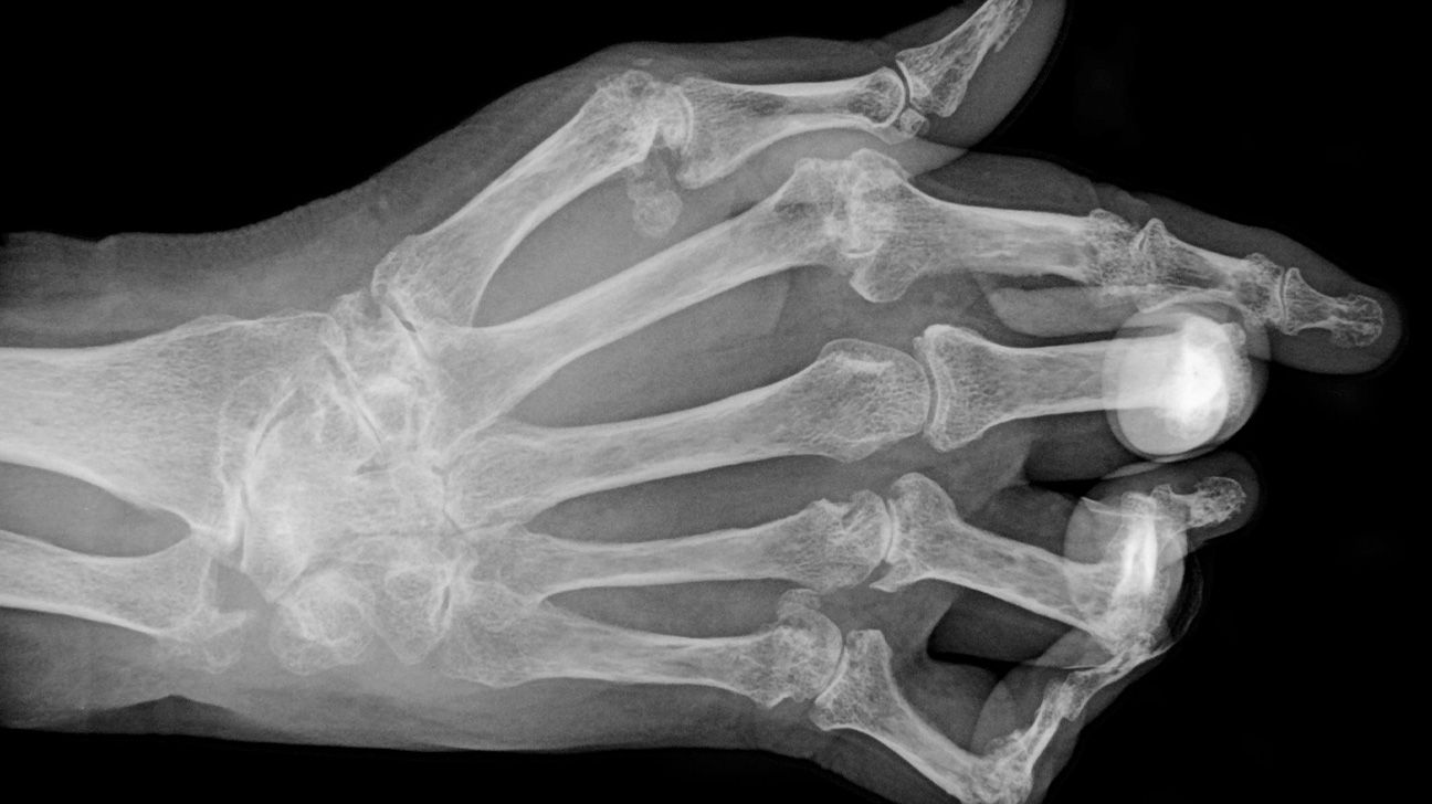
An X-ray machine uses electromagnetic radiation to create images of the inside of the body.
X-rays can show bone damage where the bone meets the joint. In particular, the X-ray can display erosion of the cortical bone, or compact bone, which is the harder layer on the outside of the bone. Cortical bone erosion is a main feature of erosive rheumatoid arthritis.
However, bone damage due to inflammation can occur gradually over time. Therefore, an X-ray will only be able to detect early rheumatoid arthritis in cases where the damage occurs quickly.
According to a
Learn about diagnosing hip arthritis with X-rays.
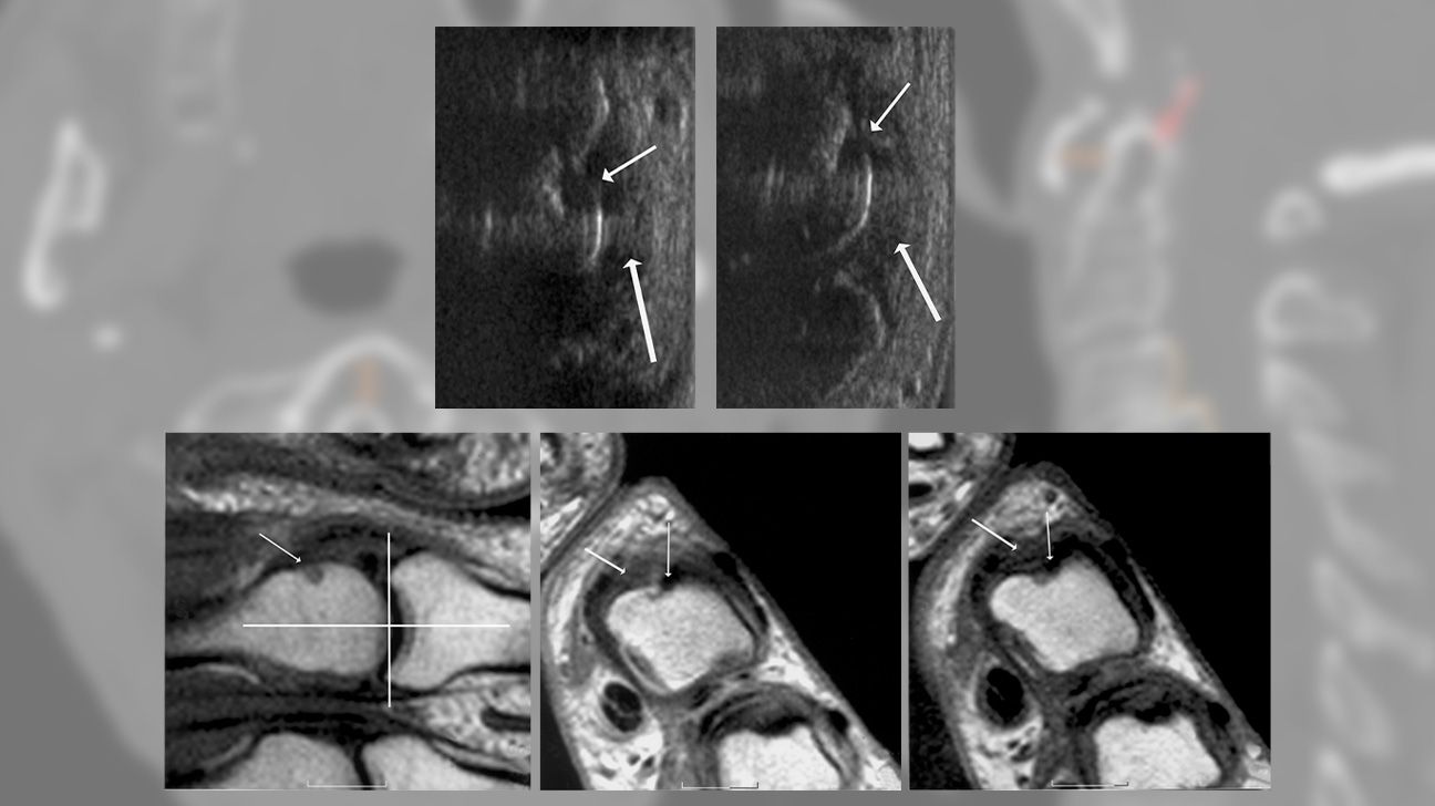
MRI uses radio waves and a powerful magnetic field to create 3D images of the whole body. It can help diagnose rheumatoid arthritis by showing any changes to the bone or cartilage.
According to the Arthritis Foundation, MRI can indicate inflammation and erosion of the bone that may not be visible on an X-ray.
MRI can also help predict the progression of the condition and monitor how effective treatments are.
Learn more about what arthritis looks like on an MRI.
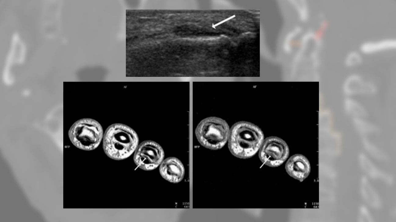
Like MRI, an ultrasound can create images of the whole body. It does so using high-frequency sound waves.
According to a 2017 article, an ultrasound can help with:
- assessing active inflammation and any inflammatory changes in the peripheral joints
- checking for tendon tears, erosion of the cortical bone, and damage to the articular cartilage
- looking for specific signs of rheumatoid arthritis, such as:
- effusion, or fluid collecting in the hollow spaces around the joint tissues
- synovitis, or inflammation of the joint’s synovial membrane
- tenosynovitis, or inflammation of the sheath that surrounds tendons
Ultrasound can also be a more cost-effective alternative to MRI.
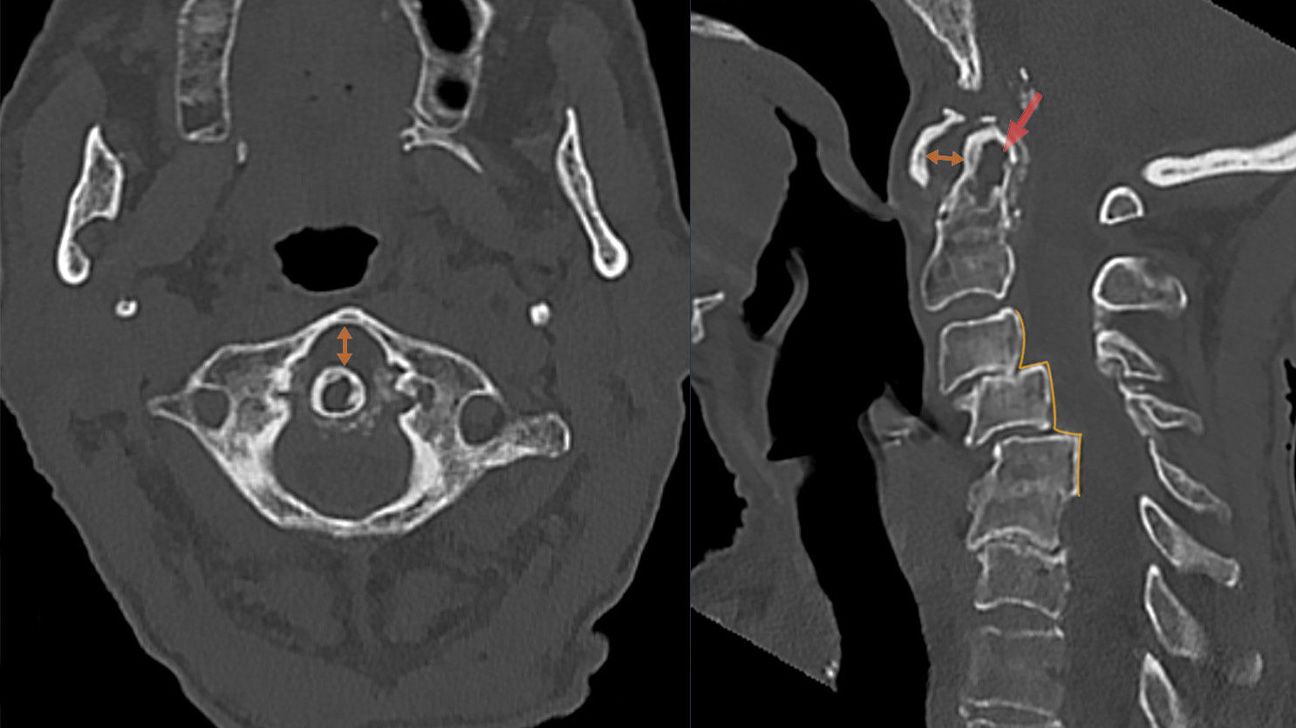
CT scans use X-rays to create detailed images of structures inside the body.
CT scans may help show early bone erosions in people with rheumatoid arthritis.
However, CT scans
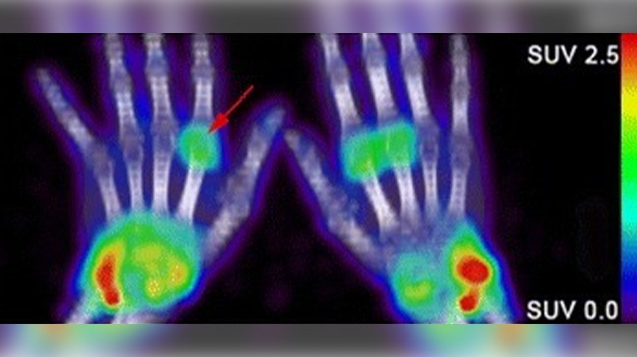
A PET scan uses radiation to show images of the inside of the body.
A combined PET-CT scan may be useful in rheumatoid arthritis to:
- diagnose the condition
- monitor how the condition responds to treatment
- predict the likelihood of remission, which refers to the period when a person has no or few symptoms
- diagnose and monitor complications
A 2022 study suggests that, while other tests may only assess certain joints, a full-body PET scan can provide a complete picture of the large and small joints simultaneously. This can help provide a better overview of the condition.
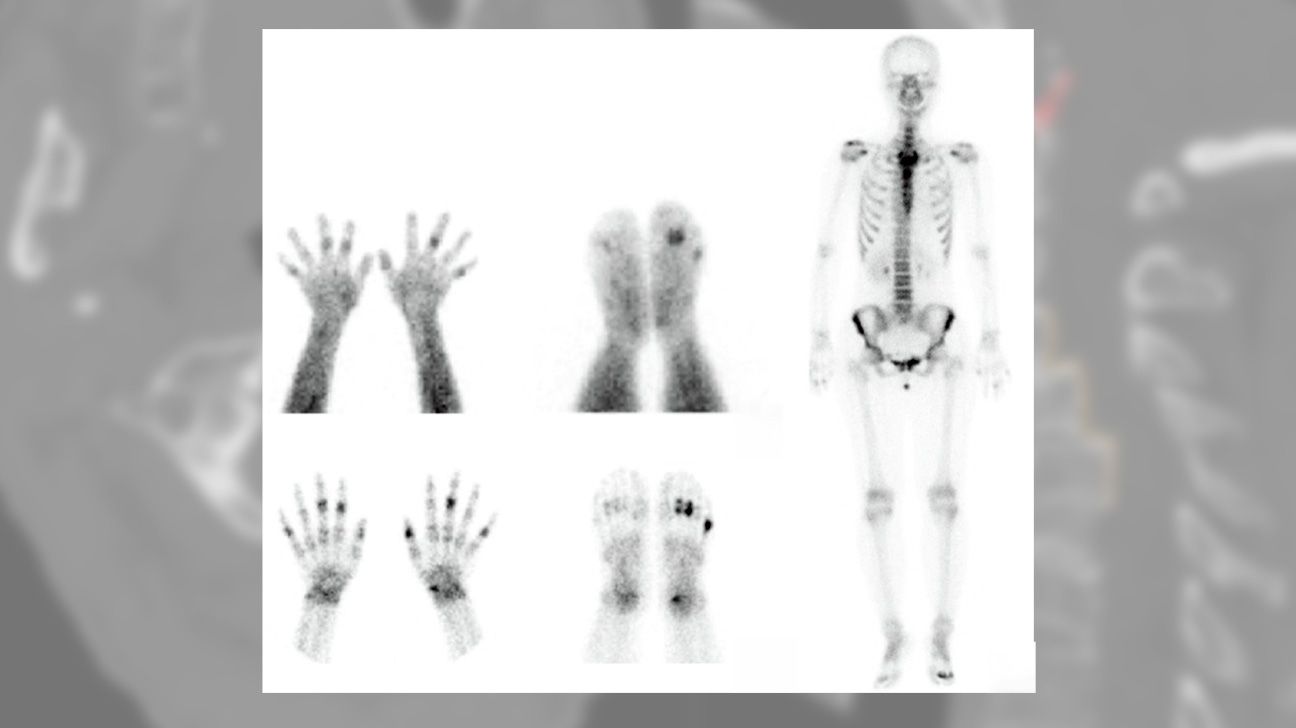
Bone scintigraphy, or a bone scan, involves injecting a radioactive tracer into the individual and then using a camera to look at the body.
A
While the research suggests that bone scans could be beneficial for detecting rheumatoid arthritis, doctors do not commonly order these tests in these situations. More research is necessary to better understand the benefits of bone scintigraphy in diagnosing and monitoring rheumatoid arthritis.
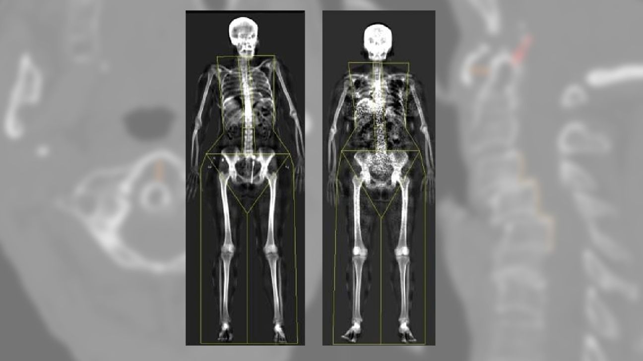
Dual-energy X-ray absorptiometry (DEXA) is an X-ray that measures bone mineral density.
Rheumatoid arthritis can lead to bone loss. DEXA can help monitor bone density in people with rheumatoid arthritis, with improvements in bone density indicating that treatment appears to be working.
People with rheumatoid arthritis are also at risk of osteoporosis. DEXA can help evaluate this risk or confirm the diagnosis.
Learn more about arthritis and bone density tests.
A doctor may order radiology or imaging tests if a person has signs or symptoms of rheumatoid arthritis. These
- inflammation
- joint pain
- joint stiffness
- fatigue
- low grade fever
- loss of appetite
If the doctor suspects rheumatoid arthritis, they will typically begin by performing a physical examination. They may then order scans and other tests.
Other tests that can help diagnose rheumatoid arthritis
- complete blood count
- rheumatoid factor (RF) blood test to check for the rheumatoid factor antibody
- anti-cyclic citrullinated peptide antibody (anti-CCP) blood test to check for anti-CCP antibodies
- erythrocyte sedimentation rate (ESR) and C-reactive protein tests to measure inflammation in the body
A person’s doctor can recommend what tests to take and answer any questions about what to expect.
Learn more about how doctors diagnose rheumatoid arthritis.
Radiology tests that can help diagnose and monitor rheumatoid arthritis include X-rays, MRIs, ultrasounds, CT scans, PET-CT scans, bone scintigraphy, and dual-energy X-ray absorptiometry (DEXA).
The imaging tests can help check for inflammation and bone erosion. DEXA measures bone density, which can help assess a person’s risk of osteoporosis.
If a doctor suspects rheumatoid arthritis, they will likely order imaging tests alongside blood tests.