Doctors use bone density testing to determine the strength of a person’s bones. These tests can show if someone has low bone density and may be at risk of osteoporosis. Doctors can use various methods to test bone density, including X-rays, CT scans, and ultrasound scans.
These tests can scan different types of bone in the body. Generally, they measure the amount of bone material in a particular section of bone, such as the hips or spine.
The more bone material a person has in their bones, the higher their bone density will be.
Read on to learn about the different bone density tests, how they work, and when a doctor might use them.
Adult humans have 206 bones, which are essential for allowing movement, protecting essential organs, and storing minerals. Bones must be dense to do these jobs.
Around
Around 10% of a person’s bone volume consists of bone cells. These produce and shape the extracellular matrix and regulate the passage of minerals in and out of the bone.
A person’s bones contain a large amount of their body’s minerals. This includes around 99% of a body’s storage of calcium, 85% of a body’s storage of phosphorus, and 40 to 60% of a body’s storage of magnesium and sodium.
Low bone density can occur due to illnesses, medications, and increasing age. When a person’s bone density lowers, they may be at greater risk of bone fractures or osteoporosis.
There are several types of bone density tests that doctors can use in clinical settings. These include the following:
- Dual energy X-ray absorptiometry (DXA): A DXA test is the
most commonTrusted Source bone density test. Doctors use these tests to determine the bone density of central bones, such as the hips or spine. - Peripheral DXA (pDXA): This is another type of DXA test. A pDXA test measures the bone density of distal bones, such as the tibia (shin bone) and radius (a bone in the forearm).
- Quantitative CT (QCT): This test measures the bone density of central bones using a CT scanner.
- Peripheral QCT: This test measures the bone density of distal bones using a CT scanner.
- Quantitative ultrasound (QUS): This type of test can show the bone density of certain distal bones, such as the tibia, radius, and calcaneus (heel bone). It does not use radiation to produce images.
All bone density tests work in similar ways. However, DXA and QCT tests use X-rays to determine bone density, while QUS tests use ultrasound.
During a DXA or QCT scan, a doctor will use a machine that can emit and detect X-rays. A person’s bones will absorb different amounts of these X-rays depending on the mineral density of their bones.
A doctor can determine how much X-ray radiation the bones absorb by comparing the amount of X-ray radiation the machine emits with the amount it detects on the other side of the body.
The doctor can then use this number to estimate a person’s bone density.
QUS scans work similarly. However, instead of emitting and detecting X-rays, they use ultrasound.
Ultrasound scans use high-frequency sound waves. These sound waves echo as they come into contact with the bones and build a picture of the inside of the body.
Unlike X-rays, ultrasound is not a form of radiation.
Some common uses for bone density tests are to detect bone fractures and conditions such as osteoporosis.
Osteoporosis is a condition that causes people to lose bone mass. It affects around
Without treatment, osteoporosis can make people particularly vulnerable to bone fractures.
Many doctors recommend that older adults have regular bone density tests because osteoporosis is more common in later life.
Healthcare professionals will give a person the results of their bone density scans in T-scores or Z-scores.
T-scores
For females in postmenopause or males ages 50 years or older, professionals will classify their bone mineral density using a T-score.
A T-score is the difference between a person’s bone mineral density and 0, which is the bone mineral density of a healthy young adult. The lower the T-score, the higher the person’s risk of a bone fracture.
The guidelines for T-scores are
- –1 or higher indicates healthy bone density
- –1 to –2.5 indicates osteopenia, a less severe form of low bone mineral density than osteoporosis
- –2.5 or lower may indicate osteoporosis
The risk of broken bones increases by 1.5 to 2 times with each 1-point drop in the T-score.
Z-scores
Z-scores are for premenopausal females and males under the age of 50. Healthcare professionals also use Z-scores to classify bone density in children.
The Z-score is the difference between a person’s bone mineral density and the average bone mineral density for healthy people of the same age, ethnicity, and sex.
If a person gets a Z-score of –2.0 or less, they have low bone mineral density. This score could indicate osteoporosis due to medications or other diseases and conditions.
A person having a bone density test does not need to do anything to prepare. They will not need to fast and may not need to undress.
However, individuals cannot wear any metallic piercings or accessories during a DXA or QCT scan, as metal can interfere with X-ray radiation.
They also
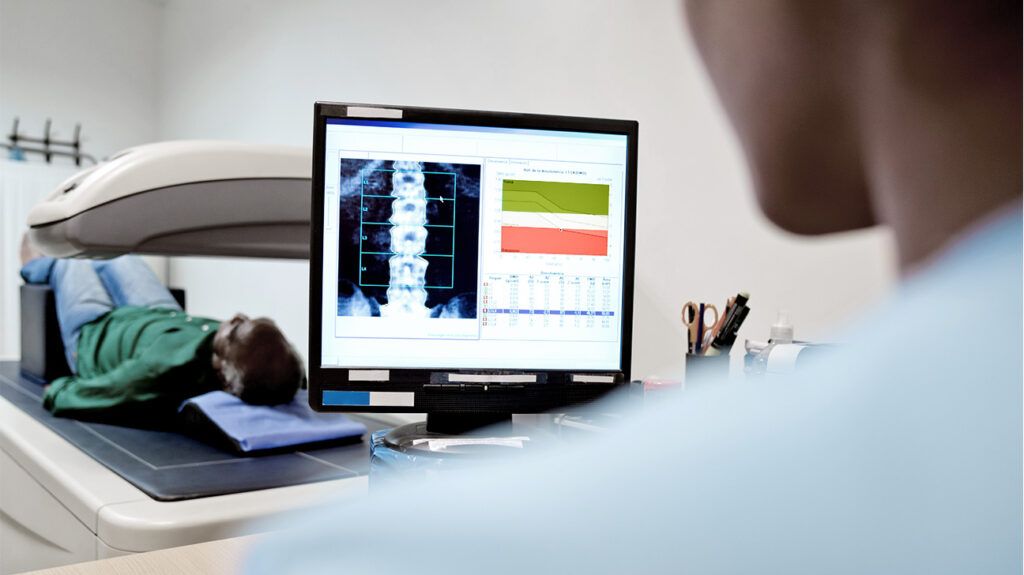
Bone density scans do not take long and generally take around 2 minutes. During the scan, a person may have to lie down on a table with their legs raised by a box.
People rarely receive their test results straight away because a trained doctor has to interpret the results first.
Different bone density tests have various benefits and limitations.
For example, DXA scans can reliably detect a person’s risk of bone fractures. However, they may be less effective for people with spinal development issues or those who have previously had spinal surgery.
QCT scans can give very precise measurements of different parts of the bone structures. However, they are less standardized than DXA tests, making it difficult to compare the results of different QCT scans.
QUS scans are helpful because the machinery is portable and does not produce harmful radiation. However, these scans may be less reliable and accurate than DXA or QCT scans.
DXT scans may also
It is not always possible for a person to improve their bone density. However, certain lifestyle changes
These lifestyle changes include:
A wide range of medications can also help people with bone density conditions such as osteoporosis.
Having sufficient bone density is important for overall health. Low bone density can be a sign of conditions such as osteoporosis and may put a person at risk of bone fractures.
Scientists have developed a wide variety of bone density testing methods. These can help doctors detect low bone density before it becomes severe or detect osteoporosis so that a person can begin treatment.
X-ray scans, CT scans, and ultrasound scans can measure a person’s bone density. Doctors use dual energy X-ray absorptiometry (DXA) scans most widely.
There are benefits and limitations to each of the different bone density tests. For instance, X-ray and CT scans use radiation, but ultrasound scans may not be as accurate.
As people age, they may need more frequent bone density tests to help ensure their bones are healthy.